Serology: It’s in the Blood — The Mad Carpenter — Crime Library
The analysis of the properties and effects of serums (blood, semen, saliva, sweat, or fecal matter) is called serology. We’ll concentrate here on the principal tests used to identify blood. According to Henry C. Lee, a forensics expert who has assisted law enforcement in over 6,000 major criminal investigations — including that of O. J. Simpson — blood evidence is found most often in “crimes of violence such as homicide, assault, and sexual assault.” It may be in the form of fresh liquid, coagulated, dried, or as a small drop or stain, and each form involves a different method of preservation and collection.
We all have about ten pints of blood getting pumped throughout our bodies. When wounded, bodies leak or spray blood, and the behavior of blood in flight tends to be unaffected by such things as temperature, humidity, or atmospheric pressure. In other words, it’s uniform.
Despite how well the crime scene may get cleaned up, even the finest trace of blood can often be detected and further tested. It is often the case that while the perpetrator may scrub down the obvious places, he can still miss between floorboards, under pipes, and inside drains. Merely by pouring water on some tiles at a murder scene and pulling them up wherever the water flowed beneath them, one detective found the only existing trace of the crime — blood. His discovery so surprised the killer, who felt certain he’d done a through job of cleaning up, that he instantly confessed.
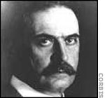
Different blood types were recognized in 1875, but it wasn’t until 1901 that Karl Landsteiner named and standardized the groups. Red blood cells carry a substance called an antigen, which produces antibodies to fight infection, and there appeared to Landsteiner to be several different types. In a centrifuge, he separated the red blood cells from the plasma or watery serum in which they are carried through the body. Then adding red blood cells from various other subjects, he found two distinct reactions — clumping and repelling. He labeled them types A (antigen A present, anti-B antibody present, but antigen B absent) and B (antigen B present, antigen A absent). Then a third reaction was labeled C (both antigens A and B absent), but was relabeled later as O. Then another type of serum was discovered, and this fourth type was labeled AB (both antigens present). It soon became clear that the blood type depended on genetic inheritance from parents, which helped with paternity tests. Type A and O are the most common in the human population, with AB the most rare.
Then Dr. Leon Lattes in Italy developed a procedure to apply blood testing to stains on fabric and other materials. He did this by finding a way to use saline solution to restore dried blood to its liquid form, although in 1932, he invented a way to test for antibodies in dried blood flakes as well.
In 1940, Landsteiner also discovered the rhesus factor in blood, labeling it Rh+ if the antigen was present in the red blood cells and Rh- if not. Today, blood typing also includes different types of enzymes and proteins that perform specific activities in the body, which helps to individualize the blood. (More than 150 serum proteins and 250 cellular enzymes have been isolated, as well as many more antigens.)
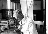
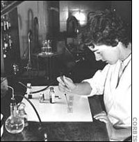
working on blood samples
Nine years went by before British scientists came to the conclusion that the nuclei of female blood cells contain a chromosome-related structure that set them apart from those of males, and they named this the Barr Body. It added one more dimension to identification via blood samples.
When a darkish substance is found at a crime scene, it must first be determined to be blood. There are several tests — presumptive tests used strictly for screening — that will differentiate between blood and other substances, but if other chemicals are present at the scene to which the test chemicals are sensitive, the tests may be vulnerable to corruption. For that reason, these tests are done with great care. A positive result from any of them is an indication to go ahead and use other tests to confirm.
Before doing anything, the crime scene investigators must take some precautions in order to avoid both biohazard to themselves and sample corruption. In his Physical Evidence in Forensic Science, Henry Lee suggests the following:
- Wear latex gloves, surgical masks, and full coverage gowns
- Eye-coverings are needed for collecting liquid samples
- Keep hands out of areas that are hidden
- Label all blood samples
- Package dry samples in bags, as well as stained clothing
- Add a note of precaution if biohazards like AIDS or hepatitis are suspected
- Decontaminate all nondisposable items
- Destroy tags, forms, or reports splashed with blood
- Clean up hands with diluted bleach, and dispose of contaminated clothing
Presumptive tests:
The first test is simply the use of a powerful light moved across every surface of a crime scene. That yields possible traces for visual inspection.
If nothing is seen, but there is reason to suspect blood had been present, a chemical called luminol is sprayed across the scene because it reacts to blood by making it luminescent. It only takes about five seconds. The procedure requires that the room be considerably darkened in order to see the faint bluish glow, and the intensity of the glow increases proportionately to the amount of blood present. It works even with old blood or diluted stains, and can illuminate smear marks where blood has been wiped away. However, there is one problem with this test: luminol can destroy the properties of the blood that investigators need for further testing. Its use is limited to proving that blood is present even if not visible.
The Kastle-Meyer Color Test uses a solution of phenolphthalein and hydrogen peroxide on a piece of filter paper, and when blood of any quantity is present, it turns pink. However, it also turns pink in the presence of potatoes or horseradish, so care must be taken at the scene.
Sometimes microcrystalline tests are also performed. The two most often used are the Takayama and Teichmann tests. Both add specific chemicals to the blood to make it form crystals with hemoglobin derivatives. These tests are also sensitive to other materials that may be present in a bloodstain.
Further testing:
From there, investigators use the precipitin test to determine whether the blood is of animal or human origin. German biologist Paul Uhlenhuth discovered that if he injected protein from a chicken egg into a rabbit, and then mixed serum from the rabbit with egg white, the egg proteins separated from the liquid to form a cloudy substance known as precipitin. In other words, it forms an antibody. In the forensic test for human blood, either a sample of the suspect blood is put into a test tube over the rabbit serum or it’s used in the “gel diffusion” test, where it’s placed in gel on a glass slide next to a sample of the reagent (anti-human serum). Passing an electric current through the glass, the protein molecules filter into the gelatin and toward each other. If a line forms where they meet — called a precipitin line — that means the sample is human blood.
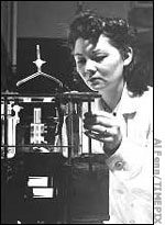
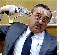
electrophoresis apparatus
After that, analysts can go ahead and determine blood type with an ABO test, and then work on the gender of the person from whom the blood came. To get a more thorough enzyme/protein profile, they use electrophoresis (a blood-soaked piece of cotton placed in gelatin on a slide and submitted to electric current).
In 1925, another blood-related discovery important to criminal investigation was made. Around 80 percent of the human population were found to be “secretors,” which means that the specific types of antigens, proteins, antibodies, and enzyme characteristic of their blood can be found in other bodily fluids and tissues. In the case of a secretor, investigators can tell the blood type by examining the saliva, teardrops, skin tissue, urine, or semen. In a rape case, for example, where the perpetrator is a secretor, potential suspects can be narrowed down through blood type analysis.
These days, thanks to discoveries in 1985, DNA technology has replaced the tests for specific enzymes and proteins. It’s more accurate to match DNA from a blood sample at a crime scene to a source than to draw up an entire blood profile.
Yet blood at a crime scene can offer even more clues than gender and type. Aside from the mere presence of blood, the different ways that blood lands on a surface has given rise to a forensic subspecialty known as blood-pattern analysis, or BPA. Let’s hear about that from one of the experts.
Serology: It’s in the Blood
Blood Pattern Analysis
Blood pattern analysis plays an important role in the reconstruction of many crime scenes. For example, when a prominent Cincinnati physician appeared to be the victim of an apparent suicide, the spatter pattern on his hand and on the couch on which he lay told a story of murder instead. The various types of bloodstains indicate how the blood was projected from the body via several factors:
- Type of injuries
- The order in which the wounds were received
- Whose blood is present
- The type of weapon that caused the injuries
- Whether the victim was in motion or lying still when the injury was inflicted
- Whether the victim was moved after the injury was inflicted
- How far the blood drops fell before hitting the surface where they were found
Blood may be dripped out, sprayed from an artery, oozed out through a large wound, or flung off a weapon raised to strike another blow. In the 1930s, Scottish pathologist John Glaister classified blood splashes into six distinct types:
- Drops on a horizontal surface
- Splashes, from blood flying through the air and hitting a surface at an angle
- Pools around the body, which can show if it’s been dragged
- Spurts from a major artery or vein
- Smears left by movement of a bleeding person
Trails, either in form of smears when a bleeding body is dragged, or in droplets when it is carried. (Trails also form when a person is wounded and walks away but leaves blood along the way.)
Any of these can be traced back to their converging point by considering such factors as the surface on which it fell, the angle it hit, and the distance it traveled.
Brian Kennedy, a sergeant with the Sacramento County Sheriff’s Department in California, is an expert in crime scene reconstruction, specializing since 1984 in bloodstain pattern analysis. For more than a decade, he has been teaching this technique to other forensic investigators and he believes that gaining insight from bloodstain patterns can strengthen interrogation strategies and provide juries with a clear visual format.
“Bloodstain patterns,” Kennedy says, “will help the investigators understand the positions and the means by which the victim and suspect moved, interacted, and struggled through the crime scene. With an understanding of what and how things occurred, investigators can focus and find fingerprints, footprints, hairs, fibers and other forms of trace evidence. The assessment of bloodstain patterns will also limit the need to collect an overabundance of redundant blood sample for DNA. Furthermore, a reconstruction of the scene helps the investigators determine which of the witnesses and suspects is telling the truth or lying.”
The shape of the blood drop itself, according to Kennedy, can reveal significant information. “The proportions of the drops can reveal the energy needed to disburse it in those dimensions. The shape of the stain can illustrate the direction in which it was traveling and angle at which it struck the surface. Choosing several stains, and using basic trigonometric functions, enables us to do a three dimensional recreation of the area of origin from which a blood-letting event occurred.”
Kennedy goes on to point out that the disruption of a blood drop on impact with a surface is directly related to the texture of the surface. “A smooth surface, such as glass,
will provide the recording of a stain with clean edges and shapes of proper geometric proportion. A rough surface, like concrete, will break the surface tension irregularly and generate a star burst, or spinning effect. An experienced analyst is able to use some of the most disrupted stains to recreate the event. Arterial spurts, for example, when compared with the anatomical location of the injury may provide information about the position when the injury was inflicted and any subsequent movement by the injured party. Castoff patterns, or drops that are thrown off of a swinging instrument in the arc of the swing, can illustrate the position of the assailant when swinging a knife, or club.”
Other crime scene analysts make the analysis of blood drops and spatters a bit more standard, but are aware, like Kennedy, that the surface type has a significant effect on just how the blood will appear. Distance estimates such as those provided in the following list are of little value unless the effects of a surface are known.
According to the basic teaching texts, the shape of a blood drop can reveal a lot about the conditions in which it fell. Given the many variations in what can happen at a crime scene, the experts don’t necessarily all agree, but a flexible rule of thumb with a generally smooth and non-porous surface might be the following:
- If blood falls a short distance — around twelve inches — at a 45-degree angle, the marks tend to be circular
- If blood drops fall several feet straight down, the edges may become crenellated, and the farther the distance from the source to the surface, the more pronounced the crenellation
- A height of six feet or more can produce small spurts that radiate out from the main drop
- If there are many drops less than an eighth of an inch across, with no larger drop, then it may be concluded that the blood spatter probably resulted from an impact
- If the source was in motion when the blood leaked or spurted, or if the drops flew through the air and hit an angled surface, the drops generally look like stretched-out exclamation marks. The end of the stain that has the smallest size blob indicates the direction in which the source was moving.
It must be emphasized that blood pattern analysis is a complicated discipline and requires experience with many different situations to learn to do an accurate reading. While any of the above statements may be true, there can also be exceptions, and all interpretations are contingent on the factors that make up the context of the crime scene, most specifically the surface on which the blood made an impact.
“All classifications of bloodstain patterns help in the reconstruction of the events,” Kennedy points out. “Spatter patterns give the nature of the force and positioning of the victim when shot or bludgeoned. Castoff patterns reveal the positioning and the possible size of the assailant. One also gets an indication of the size of the instrument swung and whether the swinger is left- or right-handed. Transfer patterns and hemorrhage or drip patterns give the direction of movement after blood is shed and can give an indication of timeframes. Arterial spurting can give the position, movement and seriousness of the injury, while ‘shadows’ — the absence of blood where one would expect to find it — suggest movement or removal of objects and changes to the scene.”
A case in England in 1984 shows the importance of blood pattern analysis. A village womanizer named Graham Backhouse was found injured in his home with slashes across his face and chest. A neighbor with whom he’d had a dispute lay dead nearby, shot by Backhouse, who claimed the man had attacked him. However, the blood patterns showed that Backhouse had been standing still or moving slowly when he was wounded, rather than being in the sort of struggle he described. In that case, the blood would have been flung against surfaces to produce the elliptical pattern. Also, there was no blood from Backhouse on his gun or near the victim. The conclusion was that he shot his victim and then inflicted the wounds on himself. Along with other evidence, the blood analysis helped to convict him of murder.
“I have worked many cases in which bloodstain patterns contributed significantly to a guilty plea or guilty verdict,” Kennedy recalls. “I have also worked cases in which the bloodstains did not support the initial allegations and the defendant was convicted of a lesser crime, or the charges were dismissed. For example in People vs. Brett Brooks Harris in Sacramento, the defendant claimed his stepfather had beaten his mother to death and then attacked him. Brett said he took the maddux handle from his stepfather and killed him in self-defense. Yet the bloodstain patterns showed that the stepfather was down and incapacitated before the mother, and both victims had been beaten more than they would have been during a single event. Based on bloodstain pattern analysis, Brett was held to answer from the preliminary hearing and, after serology validated the analysis, he pled guilty before trial.”
In another case on which Kennedy worked, People vs. Pallermo in San Joaquin County, the defendant claimed the female victim had accidentally shot herself. “However, the bloodstain pattern analysis and computer models showed the defendant’s rendition to have been an impossibility, and it placed the shooter across the room with the victim crawling into the corner to get away from the threat of the gun. The jury found the defendant guilty of second degree murder.”
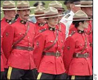
Asked about current research that may provide breakthroughs for forensic investigation, Kennedy said, “Bloodstain pattern analysis involves a constant need for research and development. The more cases that are worked and the more investigators involved, the more sophisticated the discipline will become. Probably the most advanced research has been done by members of the Crime Scene Bloodstain Section of the Royal Canadian Mount Police, Forensic Support Services. The RCMP is the only agency I know in the world that assigns and trains experienced crime scene investigators to do bloodstain pattern analysis only. They have developed a number of protocols that have been adopted around the world. Dr. Fred Carter, a professor of physics, has worked with the RCMP to develop several computer programs to teach the new analyst and help the experienced investigator.”
No matter what kind of analysis is used on the blood at a crime scene, care must be taken to handle it properly and to prevent putrefaction. Photos and notes should be taken before any blood is lifted. Samples should not be exposed to heat, moisture, or bacterial contamination, because this shortens the survival time of proteins, enzymes, and antigens. Delays in getting samples to the lab must be avoided at all cost, because it can diminish evidential value.
This was one of the central issues in the most prominent case of the past decade in which blood analysis was a primary feature: the O. J. Simpson investigation.
Since 1985, with Alec Jeffrey’s discovery of the uniqueness of portions of the DNA structure of certain genes, investigations involving blood have taken an entirely new turn. While the ultimate goal of the analysis of proteins and enzymes was to individualize blood, that’s pretty much established with DNA technology. Within a year of the discovery, DNA typing was being put to the test in criminal cases. It not only cleared one man who had confessed to a crime, but also led to the conviction of the actual killer in the same crime.
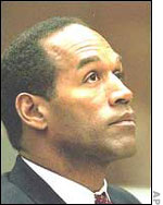
DNA can narrow down suspects in a hurry, but it’s not fool-proof. It can be challenged in court on the basis of sloppy evidence collection and the corruption of samples during testing. That was the tactic that O. J. Simpson’s defense team used to win for him an acquittal in his double murder trial. Just how did they manage to accomplish this? To trace their strategy, let’s look at the case.
On the night of July 12, 1994, Nicole Brown Simpson and Ronald Goldman were slaughtered outside her Brentwood, California home. Nicole was the former wife of football celebrity O. J. Simpson, and he was called in from out of town for questioning. Going to his home on the night of the murder, detectives had noted a bloodstain on the door of his white Ford Bronco and a trail of blood leading up to the house. That was suspicious enough to start asking questions.
When Simpson returned to Los Angeles, investigators noticed a cut on a finger of his left hand. He told several conflicting stories about how he had gotten it, which boxed him in later when blood at the crime scene indicated that the killer had been cut on his left hand and had trailed blood outside the gates. That hardly seemed coincidental.
Then when several droplets of blood at the scene failed to show a match with either of the victim’s blood types, Simpson’s blood was drawn for testing (after the droplets had already been collected). Comparison between his DNA and that of the blood at the scene showed strong similarities. The tests indicated that the drops had three factors in common with Simpson’s blood and only one person in 57 billion could produce an equivalent match. In addition, the blood was found near footprints made by a rare and expensive type of shoe — shoes that O. J. wore and that proved to be his size.
Next to the bodies was a bloodstained black leather glove that bore traces of fiber from Goldman’s jeans. The glove’s mate, stained with blood that matched Simpson’s, was found on his property. There were also traces of the blood of both victims lifted from inside Simpson’s car and house, along with blood that contained his DNA. In fact, his blood and Goldman’s were found together on the car’s console.
Forensic serologists at the California Department of Justice, along with a private contractor, did the DNA testing. Then other evidence emerged, such as the testimony of the limousine driver who came to pick Simpson up for the ride to the airport: On the night of the murder, while he waited for Simpson, he had seen a black man cross the driveway and go into the house. Then Simpson claimed that the driver had been unable to get him on the intercom because he had “overslept.” So then who was the black man who had entered the house?
When arraigned, Simpson pleaded Not Guilty and hired a defense team of celebrity lawyers. Barry Scheck and Peter Neufeld from New York were the DNA experts, renowned for their work on the Innocence Project, which used DNA analysis to defend the falsely accused. Scheck felt confident that they could produce challenges before the jury that would both educate and persuade them.
The reliability of this evidence came to be called the “DNA Wars,” and three different crime labs performed the analysis. All three determined that the DNA in the drops of blood at the scene matched Simpson’s. It was a 1 in 170 million match, using one type of analysis known as RFLP, and 1 in 240 million match using the PCR test.
Nevertheless, criminologist Dr. Henry Lee testified that there appeared to be something wrong with the way the blood was packaged, leading the defense to propose that the multiple samples had been switched. They also claimed that the blood had been severely degraded by being stored in a lab truck, but the prosecution’s DNA expert, Harlan Levy, said that the degradation would not have been sufficient to prevent accurate DNA analysis. He also pointed out that control samples were used that would have shown any such contamination, but Scheck suggested that the control samples had been mishandled by the lab — all five of them — and the jury bought it.
The evidence was damning, but the defense team managed to refocus the jury’s attention on the corruption in the Los Angeles Police Department. They then disputed the good reputation of the forensics labs, insisting that the evidence had been carelessly handled. Deliberating less than four hours, the jury freed Simpson with a Not Guilty verdict. They simply failed to understand how damning the DNA evidence really was and how ill-fitting was the defense’s logic about certain aspects of the blood at the crime scene.
Nevertheless, it can certainly be the case that what appears to be overwhelming blood analysis evidence still fails to tell the whole story. We can see that in the next case, a family tragedy that happened in Australia.
It should be kept in mind that analysis always involves interpretation. In the case below, an interpretation of the bloodstain evidence helped to convict a woman and stood up to two appeals, but turned out to have been in error.
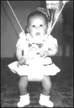
weeks, victim
In Australia in 1980, Lindy and Michael Chamberlain took their three children camping near Ayers Rock. The youngest was nine-week-old Azaria.
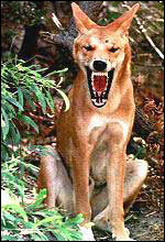
One evening, according to Lindy and Michael, they were preparing dinner at the camp barbecue site when they heard a sudden sharp cry from the tent in which Azaria was sleeping. Lindy went to check and saw a dingo, or wild dog, backing out, shaking something large in its jaws. It ran away and that’s when Lindy discovered that Azaria was gone. The dog had taken her!
Trackers searched the area to no avail. There was no sign of the missing baby or the dingo, except for footprints leading to the road and beyond. The parents grieved deeply, but eventually accepted their fate as the will of God. They assumed she was dead.
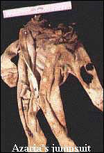
Eight days later a hiker discovered baby Azaria’s clothing in a crumpled heap west of Ayer’s Rock. Only the baby’s jacket was missing, but oddly, her undershirt was inside out and the booties were neatly laced up inside the jumpsuit. On the neck of the jumpsuit and undershirt were bloodstains that were later thought to be consistent with the type of stain that would result from a knife cut, not a bite. There were also no tooth marks on the clothing.
Around the scene, investigators found no sign of human remains, no dog hair, and no indication that violence had occurred between an animal and a baby. No dingo saliva was found on the clothing. Investigators did some experiments with caged dingoes and concluded that whatever had happened to the child had not involved a wild dog. That left human involvement — someone who left the child’s clothing several miles from where she was taken. Suspicion turned to the parents, and then more specifically to Lindy.
Lest there was doubt about whether the clothing belonged to the child, blood tests were done to determine type (no DNA testing was available then), and then compared to the Chamberlain’s blood types. The conclusion was that the clothing had belonged to Azaria. Another test showed that the undershirt had been worn the right way when the wound was made, but then someone had removed it, leaving it inside out. There also appeared to be two bloodstained prints on the jumpsuit made by the hands of a small adult, like a woman.
A search of the Chamberlain’s car produced what appeared to be the blood of an infant on the seats and on a pair of scissors in the vehicle. After that, the Chamberlains were arrested and tried for the murder of their baby daughter. They insisted they were innocent, but the evidence appeared to say otherwise. Lindy was convicted of murder and Michael was declared an accessory to the crime. Lindy went to prison.
On her behalf, many people began movements to bring out the errors made in the interpretation of evidence — particularly the experiments done with dingoes and the blood analysis. The substance found in the car, for example, was not conclusively proven by any tests to be blood. Nor was the stain on the T-shirt proven to be from a cut rather than from an arterial bleed. If the dingo had grabbed the baby by the head or neck, there would be no teeth marks on the clothing.
Then in 1986, four years after the trial, Azaria’s missing jacket was finally located — partly buried in sand near a dingo cave not far from the campsite. It was torn and bloodstained, but in good enough condition to be identified as the one Azaria wore the last time she was seen. It was sufficient for reasonable doubt and Lindy was released. The following year, the couple was officially pardoned. Not long afterward, their convictions were quashed.
No matter how sophisticated the tests, interpretation is often subject to the narrative that the investigators build, especially if the evidence is ambiguous. It can only be hoped that future technology will eliminate the gray areas and provide more conclusive proof.
Several different blood analysis techniques came together in the Caren Campano case to provide enough evidence for an arrest. She was missing and there seemed to be nothing amiss in the home… at first.
Her husband, Chris, admitted that they’d had a fight just before she had disappeared on July 1, 1992, from their Oklahoma City home. He offered to let investigators look around, which was his first mistake. A huge brownish patch on the bedroom carpet alerted them to the possibility that it was blood. They used several techniques to find out more:
1. A hema stick (microcrystal test)
This stick is coated with a blood-sensitive chemical which, when touched to a substance and then sprayed with distilled water, indicates the presence of blood. Later in the lab, they determined that it was human blood.
2. Luminol
Although the house appeared to be spotless, when this highly sensitive chemical was sprayed around the room in the dark, it illuminated so many areas that it was clear that a virtual bloodbath had taken place.
When blood flies through the air, the pattern in which it lands can determine its track, as well as the location and position of the weapon that inflicted the blow. It can also provide an estimate of how many blows were struck. Investigators found spatters on the walls, doors, and even across the ceiling. There was also a blood trail through the house and down the back outside steps. Piecing together from the splatter patterns what might have occurred, they felt certain that the victim had received numerous blows to the head with a blunt object, which collectively would have been fatal.
3. Blood Volume Test through stain recreation
On the same rug, they poured the amount of blood that would have been needed to make a stain the same size as the one they found, and then estimated that a person the size of Caren Campano would have lost at least 40 % of her blood. She could not have survived that.
4. DNA analysis – Reverse paternity test
Although Caren’s father was deceased and they had no samples of her DNA, they took blood samples from as many members of her family as they could find, hoping for a partial match with all of them. Finding it, the police had enough for an arrest on the suspicion of murder.
Finally, a year after her disappearance, they located Caren’s remains, which by this time were mostly skeletal. The dental records matched and the story told by the many fractures to the skull confirmed the theoretical scenario pieced together from the blood spatters in the Campano bedroom. Chris Campano was then convicted of the murder of his wife.
The various tests used on blood and blood patterns can offer crucial evidence in the reconstruction of a crime scene and even the proof of murder without having the body.
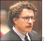
Robert Perry was found shot to death on March 29, 2001, in a back bedroom of his mobile home in Kamiah, Idaho. He was 83 and had been suffering from terminal throat cancer. Robert Perry’s 57-year-old nephew, Craig Perry, claimed to have found him while Craig’s girlfriend, Carol Flynn, then called 911 to report the suicide. The police arrived and filmed the scene for analysis, which was used when suspicions grew that this incident was no suicide.
Kamiah Police Marshal David Hasz described the scene later in court: The body lay on the floor, facing the bed. Blood was coming from the back of the frail man’s head and a .22-caliber long-barreled pistol lay near his feet. A deck of playing cards was scattered on the floor near him. Perry clutched an oxygen tube in his right hand.
Craig and Carol gave their statements and seemed quite upset, but the police had doubts when those statements seemed inconsistent with the crime scene. Their reservations deepened as the pair altered some details of their stories. Craig indicated that his uncle had made an earlier comment that he was going to “end it,” and Craig had tried to joke with him about it to dissuade him, saying he did not want to clean up the mess. They also said they’d found him on the floor, but then changed that to finding him slumped over on the bed, so close to the floor they couldn’t believe he was still sitting. Carol indicated that Craig then went to embrace the man, and finding him dead, placed him on the floor.
Prosecutors believed Craig Perry shot his uncle, and six months after the incident, Craig Perry was charged with second-degree murder.
At the trial in June 2004, the background came out. A month before the death, Craig had moved in to the trailer to care for the ailing man, and a few weeks later, Carol had joined him. At the time of the incident, both had been in the front room. Carol said she had heard a loud pop and asked Craig if he had heard it. After he asked her what she was talking about, they both heard a second one and rushed to the room.
The defense argued that Robert killed himself because he was suffering and felt like a burden to his family. Each side called experts to interpret that evidence, most specifically the wound pattern and blood spatter evidence.
The bed, complete with the same bloody sheets, was brought into the courtroom, with the blood-spattered headboard. Craig Perry’s pants were also entered into evidence, as they bore blood spatter as well.
For the prosecution, Rod Englert, a blood spatter analyst, reconstructed the event before the jury, using a stand-in person to represent Robert Perry. He said that the fine mist of blood on Craig’s pants was not a transfer smear from having handled the body but blowback from the spray released upon impact from a bullet, which meant that Craig was nearby when Robert was shot. The fact that he had blood on his pants but none on his shirt seemed to contradict his statement that he had hugged his uncle when he found him.
In addition, Englert evaluated the blood on the bed and said that the man could not have been shot while sitting there with his head slumped, because blood would have hit his legs, and there was none found in those areas. Blood found on the side of the bed indicated that Robert had been seated on the floor, leaning on the bed, when shot. Blood on Robert’s hands, Englert said, indicated that it was impossible that he had shot himself. It seemed instead to have come from contact with blood on the floor.
However, the pistol was not tested for fingerprints, and no tests were done for gunshot residue in the bedroom or on Craig Perry, so important evidence for reconstruction was lost. (A crime scene processor claimed the lab used did not accept gunshot residue tests.) Also, no second bullet was recovered from the scene, though two shell casings were.
Pathologist Dr. Michael Cihak, who performed the autopsy, demonstrated how the pistol had to have been held to make the wounds he found on the righthand side of the back of Robert’s head. He seemed to think it was unlikely that anyone would have chosen that position to shoot himself, and showed how awkward it was.
However, Cihak had already changed his own findings once, based on reports. He had located one large bullet fragment and several smaller ones in the skull, and when he learned that one casing had been found, he said he had one bullet. But when he later learned that two had been found, he changed that to say that one bullet alone could not have caused the wound he found.
Despite his findings, he admitted on cross-examination that suicide could not be ruled out.
His testimony was echoed by Dr. Vincent Di Maio, chief medical examiner from San Antonio, Texas, and nationally renowned expert on gunshot wounds. He believed that someone other than the victim had shot him, and said that only five percent of suicidal shots are behind the ear. He did not believe the man could have fired twice in succession.
To counter that, the defense called surgical pathologist George Lindholm, who testified it was possible for a man to shoot himself twice with that caliber weapon, because he might not lose consciousness the first time. Lindholm also said that, when shooting with two hands, turning the weapon upside down, it was possible to shoot oneself that way.
Stuart James, a blood spatter pattern expert from Florida, interpreted the exhibits differently. He stated that the blood on Craig’s pants could have come from the blood spewed when his uncle coughed, a frequent occurrence during the days leading up to the incident. He did not think a .22-caliber would necessarily produce that kind of blowback. In addition, expelled blood resembles high velocity impact spatter.
James had done an experiment to support his analysis. He withdrew his own blood, put it into his mouth, and coughed against the same type of material from which the suspect’s pants were made. The droplets he produced were similar in size and shape to those found on the pants. When questioned, he said that the velocity of his cough would have been similar to that of Robert Perry.
In further support of suicide, Craig claimed that he had no motive to kill his uncle. The man had no money, and Craig loved him like a father. In fact, after his own father had died, his uncle had raised him. Medical records indicated that Robert’s illness had grown worse, and his home nurse reported that he had rejected traditional cancer treatment. Two days before he died, he had coughed up blood and gone to the hospital, where doctors were unable to do anything for him.
Hours before Robert died, Carol Flynn called the hospital to report that he was having trouble breathing. That same day, she had called 911 to say he had shot himself.
Another witness said that Robert was predominantly left-handed, but had used his right hand to shoot. A neighbor and a hunting companion agreed that he was ambidextrous. Both of these witnesses had heard Robert speak about committing suicide.
Numerous character witnesses testified that Craig was a peaceful man who had loved his uncle and would not have shot anyone. He paid for all his uncle’s expenses and took care of his needs, from feeding him to getting him into bed. There was no evidence that he considered his charge a burden. A nurse said that Craig had remained positive that Robert might survive, despite the poor prognosis.
In support of the homicide theory, there was testimony that, while Robert had grown worse, he was taking simple medicines and resisting making discussions about his will. He got angry with his nurse and told her, “You already have me in the grave,” an indication that he was fierce about living. Police found a newspaper article about assisted suicide in a suitcase, along with power of attorney documents.
Also, when police had asked Craig Perry to leave the trailer on the day of the incident so they could investigate, he yelled, “You think I killed him, don’t you!” Then after being confronted with the blood analysis evidence, Craig changed his statement about hearing two pops to hearing one and then running to the bedroom — presumably to imply that he arrived in time for the second shot.
A witness also testified that when she found the second cartridge under the playing cards, Craig threw it away, saying the police must not need it.
Clearly, the crime scene was poorly processed. Overlooking a cartridge near the body and declining to do any gun shot residue analysis, which may have resolved many questions, cloud the issue.
On June 25, 2004, after two days of deliberations, the jury announced its decision: Craig Perry was acquitted of killing his uncle. Even the blood spatter evidence was ambiguous enough to allow for reasonable doubt.
Chamberlain, Lindy. Through My Eyes, Australia: William Heinemann, 1990.
Evans, Colin. The Casebook of Forensic Detection. New York: John Wiley, 1996.
Geberth, Vernon J. Practical Homicide Investigation, 3rd Ed., Boca Raton, FL: CRC Press, 1996.
Innes, Brian. Bodies of Evidence. Pleasantville, NY: Reader’s Digest Press, 2000.
Lee, Henry C and Howard A. Harris. Physical Evidence in Forensic Science. Tucson, AZ: Lawyers & Judges Publishing Company, 2000.
MacDonell, Herbert L. Bloodstain Patterns. Corning, NY: Laboratory of Forensic Science, 1993.
Nickell, Joe & John Fischer. Crime Science: Methods of Forensic Detection. Lexington, KY: The University Press of Kentucky, 1999.
Owen, David. Hidden Evidence: Forty True Crimes and How Forensic Science Helped Solve Them. Buffalo, NY: Firefly Books, 2000.
Saferstein, Richard. Criminalistics: An Introduction to Forensic Science. 5th Edition. Englewood Cliffs, NJ: Prentice Hall, 1995.
Schiller, Lawrence. American Tragedy: The Uncensored Story of What Happened Behind Closed Doors. New York: Avon, 1996.
“The House that Roared,” Forensic Files, Court TV, January 11, 2001.
Young, Norman H. Innocence Regained, The Federation Press, 1989.